Management of Patient with Limb Girdle Muscular Dystrophy for ERCP and Regional Anaesthesia for Laparoscopic Cholecystectomy: Case Report
Vol 3 | Issue 1 | January-June 2022 | Page 27-30 | Shalini Saksena , Arnab Paul
DOI: 10.13107/ijra.2022.v03i01.051
Authors: Shalini Saksena [1], Arnab Paul [1]
[1] Department of Anaesthesia, PD Hinduja National Hospital & MRC, Mahim, Mumbai, Maharashtra, India.
Address of Correspondence:
Dr. Arnab Paul,
Department of Anaesthesia, PD Hinduja National Hospital & MRC, Mahim, Mumbai, Maharashtra, India.
E-mail: dr.arnabpaul88@gmail.com
Abstract
Limb-Girdle muscular dystrophies (LGMDs) are a clinically and genetically heterogeneous group of disorders characterized in general by predominantly limb-girdle weakness. It manifests as proximal muscle weakness involving the pelvic girdle and scapular girdle. We report the anaesthetic management of a patient who is a known case of limb-girdle muscular dystrophy, presented with cholecystitis to our institution. Patient underwent endoscopic retrograde cholangio pancreatography (ERCP) under total intravenous anaesthesia followed by laparoscopic cholecystectomy under spinal anaesthesia which were managed successfully.
Keywords: Muscular Dystrophies, Limb-Girdle, Malignant Hyperthermia, Anaesthesia
References
1. Jayadev S. Muscular Dystrophies. Brenner’s Encyclopedia of Genetics 2013; 2: 522-4.
2. Allen T, Maguire S. Anaesthetic management of a woman with autosomal recessive limb-girdle muscular dystrophy for emergency caesarean section. Int J Obstet Anesth 2007; 16:370–374.
3. Iyadurai SJ, Kissel JT. The Limb-Girdle Muscular Dystrophies and the Dystrophinopathies. Continuum (Minneap Minn). 2016;22(6, Muscle and Neuromuscular Junction Disorders):1954–77
4. Chu ML, Moran E. The limb-girdle muscular dystrophies: is treatment on the horizon? Neurotherapeutics. 2018; 15:849–62.
5. Richa FC. Anaesthetic management of a patient with limb-girdle muscular dystrophy for laparoscopic cholecystectomy. Eur J Anaesthesiol 2011; 28:72–73.
6. Segura LG, Lorenz JD, Weingarten TN, et al. Anaesthesia and Duchenne or Becker muscular dystrophy: review of 117 anesthetic exposures. Paediatr Anaesth 2013; 23:855–864.
7. Kim TW, Nemergut ME. Preparation of modern Anaesthesia workstations for malignant hyperthermia-susceptible patients: a review of past and present practice. Anesthesiology. 2011;114(1):205–12.
8. Wappler F. Anaesthesia for patients with a history of malignant hyperthermia. Curr Opin Anaesthesiol. 2010;23(3):417–22.
9. Bajwa SJ, Kulshrestha A. Anaesthesia for laparoscopic surgery: General vs regional anaesthesia. J Min Access Surg 2016; 12:4-9.
10. Catani M, Guerricchio R, De Milito R, et al. ‘Low-pressure’ laparoscopic cholecystectomy in high-risk patients (ASA III and IV): our experience. Chir Ital 2004; 56:71–80.
11. Van Obbergh LJ, Corteel J, Papadopoulos J, et al. Anaesthesia for a child suffering from a deletion in the Xp21 loci resulting in Duchenne disease, glycerol kinase deficiency and congenital adrenal hypoplasia. Paediatr Anaesth 2011; 21:1085–87.
| How to Cite this Article: Saksena S, Paul A | Management of Patient With Limb Girdle Muscular Dystrophy for ERCP and Regional Anaesthesia for Laparoscopic Cholecystectomy: Case Report | International Journal of Regional Anaesthesia | January-June 2022; 3(1): 27-30.
|
(Abstract Text HTML) (Download PDF)
Dual Guidance in the Era of Ultrasound: An Overlooked Necessity or a Luxury!
Vol 3 | Issue 1 | January-June 2022 | Page 35-36 | Vedhika Shanker, Tuhin Mistry, Gurumoorthi Palanichamy, Jagannathan Balavenkatasubramanian
DOI: 10.13107/ijra.2022.v03i01.53
Authors: Vedhika Shanker [1], Tuhin Mistry [1], Gurumoorthi Palanichamy [1], Jagannathan Balavenkatasubramanian [1]
[1] Department of Anaesthesiology, Ganga Medical Centre & Hospitals Pvt Ltd, Coimbatore, India.
Address of Correspondence
Dr. Tuhin Mistry,
Department of Anaesthesiology, Ganga Medical Centre & Hospitals Pvt Ltd, Coimbatore, India.
E-mail: tm.tuhin87@gmail.com
Short Communication
Real-time ultrasonography (USG) guidance has revolutionized the practice of regional anesthesia (RA). As an adjunct to USG, nerve stimulation has been advocated for accurate and safe delivery of local anesthetic (LA) while performing peripheral nerve blocks [1]. This letter highlights the importance of dual guidance infraclavicular brachial plexus block (BPB) in a polytrauma patient for forearm surgery.
A 54-year-old male was brought to the emergency with an alleged history of a road traffic accident and multiple injuries, including left middle third clavicle fracture, bilateral multiple rib fractures, closed distal third both bones fracture of right forearm, scalp hematoma of the left parietal area, and left-sided pneumothorax. On arrival at the resuscitation bay, the overall pain score was 9/10 on a numeric rating scale. An intercostal drain was inserted, and he had been placed on noninvasive ventilation (NIV). A multimodal analgesia regimen was started, including continuous thoracic epidural, intravenous paracetamol 15 mg/kg, tramadol 2 mg/kg, and transdermal 10 mg buprenorphine patch. The patient has been on regular treatment for type 2 diabetes mellitus and hypertension for 15 years. He had suffered two episodes of myocardial infarction 8 years ago, for which he had undergone percutaneous transluminal coronary angioplasty and was on dual antiplatelet therapy. The transthoracic echocardiography revealed mild left ventricular hypertrophy, hypokinetic posterior, lateral, and inferior walls with a left ventricular ejection fraction of 40%. He also suffered an ischemic cerebrovascular accident involving the left middle cerebral artery 6 years ago. The patient had residual weakness of the right-sided hemiparesis, dysphagia, and slurring of speech. He was scheduled for open reduction and internal fixation with plating both right forearm bones 3 days after admission. The plan was to provide surgical anesthesia with a right-sided diaphragm sparing BPB. The anesthesia plan was explained to the patient and relatives, and informed written consent was obtained.
The patient was positioned supine with head-end elevation at 30° in the operation theater, and the ipsilateral arm was abducted. Standard monitors were attached, and a scout scan was performed with a high-frequency linear array transducer (Sonosite HFL 38xp/13–6 MHz; Fujifilm SonoSite Inc., Bothell, WA, USA) to assess the viability of the anesthetic plan (Fig. 1a). The right infraclavicular BPB was performed under dual guidance (USG and electrostimulatilation) with a 100 mm nerve block needle and 15 ml 0.75% ropivacaine and 4 mg dexamethasone was administered (Fig. 1b). Each cord of the brachial plexus was simulated separately, and 5 ml of LA was deposited after obtaining desired responses at <0.5 mA current, 0.1 ms impulse duration, and a frequency of 2 Hz. The lateral, posterior, and medial cords were identified by elbow flexion, wrist extension, and wrist flexion, respectively. The block was successful, and the procedure went off without any complications.
BPB above the clavicle is widely practiced for various upper limb surgeries. We ruled out this option to avoid inadvertent phrenic nerve palsy. Our patient was on intermittent NIV, and the procedure was undertaken once the patient could tolerate NIV-free periods without any respiratory distress. However, the challenge of the patient’s inability to lie supine remained. The costoclavicular approach could not be instituted as the patient had a right-sided subclavian central venous catheter. Hence, correct transducer placement and proper visualization of the brachial plexus were not possible (Fig. 1c). We also excluded the possibility of axillary BPB due to the presence of fungal skin infection. We opted for USG guided infraclavicular BPB, but discrimination of individual cords was not feasible. Hence, we used a combination of ultrasound and nerve stimulation for a sure-fire successful RA technique.
A successful infraclavicular BPB can be achieved either with electrostimulation or ultrasound guidance in experienced hands. However, USG shortens performance time compared to the dual-motor endpoint stimulation [2]. Although the LA deposition at a single point, cranioposterior to the axillary artery, could result in successful infraclavicular BPB, the success rate was reported to be higher with multiple-injection (53–100%) [4]. Gurkan et al. reported a similar success rate between dual guidance (95%) and single motor endpoint stimulation (93%) [5]. Hence, the use of ultrasound without neurostimulation may be sufficient to achieve a successful infraclavicular BPB. However, in particular cases, electrostimulation as an adjunct may help in the identification of individual cords based on the motor response as well as act as a safety monitor to prevent intraneural injection [1].
To conclude, Dual guidance was necessary for our patient to perform the infraclavicular BPB. Ultrasound helped in real-time visualization of spread and reduced the LA volume, while peripheral nerve stimulation aided in accurate localization of cords with evoked motor responses.
References
1. Gadsden JC. The role of peripheral nerve stimulation in the era of ultrasound-guided regional anaesthesia. Anaesthesia 2021;76 Suppl 1:65-73.
2. Brull R, Lupu M, Perlas A, Chan VW, McCartney CJ. Compared with dual nerve stimulation, ultrasound guidance shortens the time for infraclavicular block performance. Can J Anaesth 2009;56:812-8.
3. Dingemans E, Williams SR, Arcand G, Chouinard P, Harris P, Ruel M, et al. Neurostimulation in ultrasound-guided infraclavicular block: A prospective randomized trial. Anesth Analg 2007;104:1275-80.
4. Sauter AR, Dodgson MS, Stubhaug A, Halstensen AM, Klaastad Ø. Electrical nerve stimulation or ultrasound guidance for lateral sagittal infraclavicular blocks: A randomized, controlled, observer-blinded, comparative study. Anesth Analg 2008;106:1910-5.
5. Gurkan Y, Acar S, Solak M, Toker K. Comparison of nerve stimulation vs. ultrasound-guided lateral sagittal infraclavicular block. Acta Anaesthesiol Scand 2008;52:851-5.
| How to Cite this Article: Shanker V, Mistry M, Palanichamy G, Balavenkatasubramanian J | Dual Guidance in the Era of Ultrasound: An Overlooked Necessity or a Luxury! | International Journal of Regional Anaesthesia | January-June 2022; 3(1): 35-36.
|
(Abstract Text HTML) (Download PDF)
The Use of Stellate Ganglion Block and Interscalene Brachial Plexus Catheter to Treat Shoulder Hand Syndrome – A Case Report
Vol 3 | Issue 1 | January-June 2022 | Page 23-26 | Amitav V. Philip, Kiran Sasi, Rahul Pillai, Sajan Philip George
DOI: 10.13107/ijra.2022.v03i01.050
Authors: Amitav V Philip [1], Kiran Sasi [2], Rahul Pillai [1], Sajan Philip George [1]
[1] Department of Anaesthesia, Christian Medical College, Vellore, Tamil Nadu, India
[2] Department of Hand and Leprosy Reconstructive Surgery, Paul Brand Centre, Christian Medical College, Vellore, Tamil Nadu, India.
Address of Correspondence
Dr. Amitav V Philip,
Assistant Professor, Department of Anaesthesia, Christian Medical College, Vellore, Tamil Nadu, India.
E-mail: a.philip442@gmail.com
Abstract
Complex regional pain syndrome (CRPS) may develop following trivial trauma and can lead to chronic debilitating pain and dysfunction. There is limited use of sympathetic blocks in the management of CRPS; however, in patients with sympathetically mediated pain, this modality may be of value. In this paper, we report the case of a 50-year-old milkman who was diagnosed with left sided Shoulder Hand Syndrome (CRPS type 1) following prolonged immobilization after sustaining a left 3rd metacarpal fracture. The management plan included the use of a single injection stellate ganglion block and an interscalene brachial plexus catheter which was maintained for one week in order to mitigate the pain cycle. The catheter was maintained for 7 days which improved the compliance to physical therapy and psychosocial rehabilitation of the patient. The patient reported excellent pain relief with return to his work.
Keywords: CRPS type 1, Interscalene brachial plexus catheter, Stellate ganglion block
References
1. Birklein F, Dimova V. Complex regional pain syndrome–up-to-date. PAIN Rep. 2017 Dec;2(6):e624.
2. Detaille V, Busnel F, Ravary H, Jacquot A, Katz D, Allano G. Use of continuous interscalene brachial plexus block and rehabilitation to treat complex regional pain syndrome of the shoulder. Ann Phys Rehabil Med. 2010 Aug;53(6–7):406–16.
3. Ferrillo MG. Treatment of complex regional pain syndrome with stellate ganglion local anesthetic blockade: a case report of one patient’s experiences with traditional bupivacaine HCl and liposome bupivacaine. Clin Case Rep. 2016 Jul 27;4(9):861–5.
4. Bruehl S. Complex regional pain syndrome. BMJ. 2015 Jul 29;h2730.
5. Bruehl S, Warner DS. An Update on the Pathophysiology of Complex Regional Pain Syndrome. Anesthesiology. 2010 Sep 1;113(3):713–25.
6. Ganty P, Chawla R. Complex regional pain syndrome: recent updates. Contin Educ Anaesth Crit Care Pain. 2014 Apr 1;14(2):79–84.
7. Albazaz R, Wong YT, Homer-Vanniasinkam S. Complex Regional Pain Syndrome: A Review. Ann Vasc Surg. 2008 Mar 1;22(2):297–306.
8. Cepeda MS, Lau J, Carr DB. Defining the Therapeutic Role of Local Anesthetic Sympathetic Blockade in Complex Regional Pain Syndrome: A Narrative and Systematic Review. Clin J Pain. 2002 Aug;18(4):216–33.
9. Yucel I, Demiraran Y, Ozturan K, Degirmenci E. Complex regional pain syndrome type I: efficacy of stellate ganglion blockade. J Orthop Traumatol Off J Ital Soc Orthop Traumatol. 2009 Dec;10(4):179.
10. Schürmann M, Gradl G, Wizgal I, Tutic M, Moser C, Azad S, et al. Clinical and Physiologic Evaluation of Stellate Ganglion Blockade for Complex Regional Pain Syndrome Type I. Clin J Pain. 2001 Mar;17(1):94–100.
11. Lierz P, Hoffmann P, Felleiter P, Hörauf K. [Interscalene plexus block for mobilizing chronic shoulder stiffness]. Wien Klin Wochenschr. 1998 Nov 13;110(21):766–9.
12. Detaille V, Busnel F, Ravary H, Jacquot A, Katz D, Allano G. Use of continuous interscalene brachial plexus block and rehabilitation to treat complex regional pain syndrome of the shoulder. Ann Phys Rehabil Med. 2010 Sep;53(6–7):406–16.
| How to Cite this Article: Philip AV, Sasi K, Pillai R, George SP | The Use of Stellate Ganglion Block and Interscalene Brachial Plexus Catheter to Treat Shoulder Hand Syndrome – A Case Report | January-June 2022; 3(1): 23-26.
|
(Abstract Text HTML) (Download PDF)
IJRA | Volume 2 | Issue 2 | July-December 2021
Click on the Page
Regional Anaesthesia to the Rescue: A Post-Covid Patient with Lumbar Spine Injury Undergoing Lower Limb Orthopedic Surgery
Vol 2 | Issue 2 | July-December 2021 | Page 141-142 | Vandana Mangal, Hardika Mangal, Aashna Pareek, Tuhin Mistry
DOI: 10.13107/ijra.2021.v02i02.043
Authors: Vandana Mangal [1], Hardika Mangal [1], Aashna Pareek [1], Tuhin Mistry [2]
[1] Department of Anaesthesiology, Trauma Centre, S. M. S. Medical College, Jaipur, India.
[2] Department of Anaesthesiology, Ganga Medical Centre & Hospitals Pvt Ltd, Coimbatore, India.
Address of Correspondence
Dr Tuhin Mistry,
Department of Anaesthesiology, Ganga Medical Centre & Hospitals Pvt Ltd, Coimbatore, India.
E-mail: tm.tuhin87@gmail.com
Letter to Editor
Sir,
Coronavirus disease of 2019 (COVID-19) has revamped the management of trauma patients worldwide like many other specialties. Regional anaesthesia (RA) techniques such as central neuraxial block (CNB) and/or peripheral nerve block (PNB) have been advocated strongly over general anesthesia (GA) whenever feasible by various societies [1]. RA not only avoids aerosol generation in COVID-19 patients, but it may also reduce postoperative pulmonary complications, especially in covid recovered patients with compromised lung function. We wish to share our experience using peripheral nerve blocks as an alternative to central neuraxial blocks for managing lower limb fracture in a post-covid patient with lumbar spine injury.
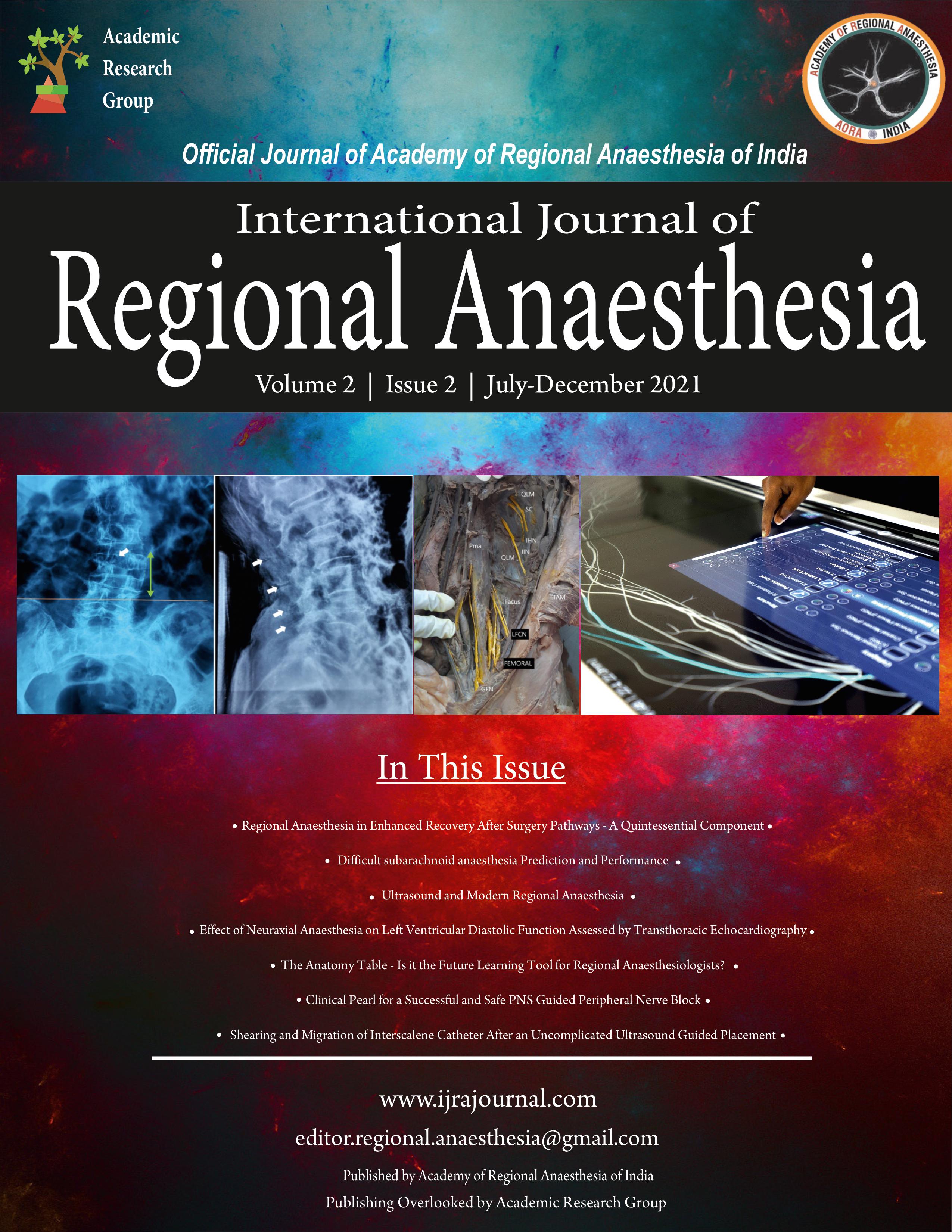
A 33-years-old lady was brought to the emergency room with an alleged history of severe back pain and bilateral leg injury following a fall from height. On examination, her heart rate, blood pressure, and room air saturation were 118 beats/min, 90/70 mm Hg, and 56%, respectively. Following primary resuscitation, she was put on a non-rebreather mask with 15 L of oxygen. Radiological investigations revealed fractures of bilateral leg bones (both the tibia and fibula) and L1vertebral body (AO type A2) (Figure 1a). High-resolution computed tomography of the chest showed bilateral ground-glass opacities and a severity score of 18/25 (Figure 1b). Her Reverse Transcription Polymerase Chain Reaction test for COVID-19 was negative, but serum inflammatory markers were increased. Her injuries were managed conservatively, and the surgery was deferred. She was shifted to the intensive care unit and managed with antibiotics, steroids, anticoagulants, and noninvasive ventilation. She was gradually weaned off from oxygen support after 25 days. She was scheduled for intramedullary nailing of the left tibia and closed reduction and plaster cast application of the right leg. Her neurological assessment revealed normal sensory and medical research council grade 3 muscle powers. The anesthesia plan was discussed with the patient and her relatives and informed written consent was obtained.
In the operating room, standard monitors were attached, and one 18 G intravenous (IV) cannula was secured. The patient was placed in the supine position. Under all aseptic precautions, ultrasound-guided left-sided femoral nerve block (FNB) and popliteal sciatic nerve block (PSNB) were performed (Figure 1c,d,e,f) using a 23G 3.5 inch Quincke spinal needle and high-frequency linear transducer (L38e, 10-5 MHz, MicroMaxx, Fujifilm SonoSite Inc., Bothell, WA, USA). 10 ml of 0.5 % bupivacaine and 20 ml of 0.5% bupivacaine were administered for FNB and PSNB, respectively, after negative aspiration for blood. Intraoperatively, IV paracetamol 1 gm, ketorolac 30 mg, and 8 mg dexamethasone were given. Closed reduction and intramedullary nailing of the left tibia was completed in one hour. Then, IV fentanyl 25 µg was given, and closed reduction and cast application of the right leg was done. The patient was comfortable throughout the surgery without any significant change in hemodynamics and did not require oxygen or any additional anesthetic medication. She was under observation in the post-anesthesia care unit for the next 24 hours. She recovered well and was discharged after five days.
The CNB or PNB is the preferred RA technique as the respiratory functions are preserved. The subarachnoid block or combined spinal-epidural anesthesia is usually practiced for manipulations and fixation of lower limb fractures as it provides a dense sensory and motor blockade. We avoided CNB as our patient had a fracture of the lumbar spine. We preferred PNB over CNB or GA because of the second wave of COVID-19, superadded oxygen scarcity, and the compromised lung function following covid pneumonia. Ultrasound-guided combined PSNB and adductor canal block has been used for below-knee surgeries in high-risk patients to maintain hemodynamic stability and postoperative pain management [2]. We used a combination of FNB and PSNB for intramedullary tibial nailing in our patient. This combination is a simple and straightforward technique to avoid CNB or GA in below-knee surgeries where use of tourniquet is not necessary. However, this appears to be an underutilized RA technique as there is a paucity in the current literature. Selvi et al. used a combination of FNB and PSNB in a patient with severe emphysematous lung disease for femoral-popliteal arterial bypass surgery [3]. Imbelloni et al. used a lateral, continuous, combined FNB and high sciatic nerve block via a single skin puncture for postoperative analgesia in a supine adult patient undergoing tibial intramedullary nailing [4]. The detection of compartment syndrome may get delayed because of the insensibility of the nerves following the block. So, we objectively assess the patient every three hours for signs of compartment syndrome that did not develop.
We faced a challenge in performing PSNB as the patient was supine and could not be turned lateral or prone position due to the presence of multiple. The anterior approach to high sciatic nerve block would have been more appropriate, but the curvilinear probe was unavailable. Hence, we performed the PSNB via lateral approach in the supine position (Figure 1e,f) as described by Gray and colleagues [5]. This technique was convenient for the patient and offered optimal needle visibility.
The combination of FNB and PSNB suited our patient. More extensive studies need to be done on a combination of blocks for outpatients coming with closed fractures of leg bones in addition to high-risk patients where CNB has to be avoided.
References
1. Macfarlane AJR, Harrop-Griffiths W, Pawa A. Regional anaesthesia and COVID-19: first choice at last? Br J Anaesth. 2020;125:243-7.
2. Arjun B K, Prijith R S, Sreeraghu G M, Narendrababu M C. Ultrasound-guided popliteal sciatic and adductor canal block for below-knee surgeries in high-risk patients. Indian J Anaesth 2019; 63:635-9.
3. Selvi O, Bayserke O, Tulgar S. Use of Femoral and Sciatic Nerve Block Combination in Severe Emphysematous Lung Disease for Femoral Popliteal Arterial Bypass Surgery. Cureus. 2018;10:e2140.
4. Imbelloni L. E., Rava C., Gouveia M. A. A new, lateral, continuous, combined, femoral-sciatic nerve approach via a single skin puncture for postoperative analgesia in intramedullary tibial nail insertion. Local and Regional Anesthesia. 2013; 6:9–12.
5. Gray AT, Huczko EL, Schafhalter-Zoppoth I. Lateral popliteal nerve block with ultrasound guidance. Reg Anesth Pain Med. 2004;29:507-9.
| How to Cite this Article: Mangal V, Mangal H, Pareek A, Mistry T | Regional Anaesthesia to the Rescue: A Post- Covid Patient with Lumbar Spine Injury Undergoing Lower Limb Orthopedic Surgery | International Journal of Regional Anaesthesia | July-December 2021; 2(2): 141-142.
|
(Abstract Text HTML) (Download PDF)
Shearing and Migration of Interscalene Catheter After an Uncomplicated Ultrasound Guided Placement
Vol 2 | Issue 2 | July-December 2021 | Page 137-140 | Shashank Rane, Vijay Shetty, Sitaram Prasad, Rajat Bhargava
DOI: 10.13107/ijra.2021.v02i02.042
Authors: Shashank Rane [1], Vijay Shetty [1], Sitaram Prasad [2], Rajat Bhargava [3]
[1] Department of Anaesthesia, Fortis Hospital, Mumbai, Maharashtra, India.
[2] Department of Plastic Surgery, Fortis Hospital, Mumbai, Maharashtra, India.
[3] Department of Radiology, Fortis Hospital, Mumbai, Maharashtra, India.
Address of Correspondence
Dr. Shashank Rane,
Department of Anaesthesia, Fortis Hospital, Mumbai, Maharashtra, India.
E-mail: shankrane81@gmail.com
Abstract
We report on the case of shearing and migration of an interscalene nerve catheter in 68- year-old female who underwent a left shoulder surgery. The catheter was placed under ultrasound guidance without any apparent complications. Continuous interscalene nerve block was successfully used to complement General Anaesthesia and provide postoperative pain relief. On the second day, at the time of catheter removal, the catheter inadvertently sheared at the point of insertion with the distal 7 centimetres migrating under the skin. The axial and coronal CT scan sections confirmed the migration of the catheter posterolateral to subclavian artery in the vicinity of the Brachial plexus. Patient did not have any pain or neurological deficit. Surgery was performed to extract the catheter, which was found in brachial plexus sheath between lower end of Scalenus Anterior and Scalenus Medius.
Keywords: Interscalene catheter, Shearing, Migration
References
1. Capdevila X, Pirat P, Bringuier S, et al. Continuous peripheral nerve blocks in hospital wards after orthopedic surgery: A multicenter prospective analysis of the quality of postoperative analgesia and complications in 1,416 patients. Anesthesiology 2005;103:1035–45.
2. Ates Y, Yucesoy CA, Unlu MA, Saygin B, Akkas N. The mechanical properties of intact and traumatized epidural catheters. Anesth Analg 2000;90:393–9.
3. Hadzic A, ed. New York School of Regional Anesthesia. Textbook of Regional Anesthesia and Acute Pain Management. New York: McGraw-Hill Medical Pub. Division; 2007:412–3.
4. Despond O, Kohut GN. Broken interscalene brachial plexus catheter: surgical removal or not? Anesth Analg. 2010 Feb 1;110(2):643-4. doi: 10.1213/ane.0b013e3181c62a05. PMID: 20081147.
5. Mitra R, Fleischmann K. Management of the sheared epidural catheter: Is surgical extraction really necessary? J Clin Anesth 2007;19:310–4.
6. Bowens C Jr, Briggs ER, Malchow RJ. Brachial plexus entrapment of interscalene nerve catheter after uncomplicated ultrasound-guided placement. Pain Med. 2011;12:1117-20.
7. Ilfeld BM, Morey TE, Enneking FK. Infraclavicular perineural local anesthetic infusion: A comparison of three dosing regimens for postoperative analgesia. Anesthesiology 2004;100:395–402.
8. Brenier G, Salces A, Magues JP, Fuzier R. Peripheral nerve catheter entrapment is not always related to knotting. Can J Anaesth 2010;57:183–4.
| How to Cite this Article: Rane S, Shetty V, Prasad S, Bhargava R | Shearing and Migration of Interscalene Catheter After an Uncomplicated Ultrasound Guided Placement | International Journal of Regional Anaesthesia | July-December 2021; 2(2): 137-140.
|