Effect of Neuraxial Anaesthesia on Left Ventricular Diastolic Function Assessed by Transthoracic Echocardiography
Vol 2 | Issue 2 | July-December 2021 | Page 131-136 | Maithriye Kavishree, Srinath Damodaran, Sharanu Patil, Kumar Belani, Muralidhar Kanchi
DOI: 10.13107/ijra.2021.v02i02.041
Authors: Maithriye Kavishree [1], Srinath Damodaran [1], Sharanu Patil [2], Kumar Belani [3], Muralidhar Kanchi [1]
[1] Department of Cardiac Anaesthesia, Narayana Institute of Cardiac Sciences, Narayana Health City, Bangalore, Karnataka, India.
[2] Department of Anaesthesia and Intensive care, Sparsh Hospital, Bangalore, Karnataka, India.
[3] Department of Cardiac Anaesthesia, Masonic Children’s Hospital, University of Minnesota, Minneapolis, United States of America.
Address of Correspondence
Dr. Muralidhar Kanchi
Academic Director, Senior Consultant & Professor, Department of Cardiac Anaesthesia, Narayana Institute of Cardiac Sciences, Narayana Health City, Bangalore, Karnataka, India.
E-mail: muralidhar.kanchi.dr@narayanhealth.org
Abstract
Purpose: To evaluate the effect of neuraxial anaesthesia on left ventricular (LV) diastolic function in clinical setting using transthoracic echocardiography (TTE).
Methods: This prospective observational study was performed in 50 adult patients undergoing elective orthopaedic surgical procedures under neuraxial anaesthesia for lower limb surgery. TTE was performed before, 20, 40 and 60 minutes after neuraxial anaesthesia. Pulsed wave Doppler of the transmitral flow (TMF), pulmonary venous flow (PVF), deceleration time (DT) and propagation velocity (Vp) were measured. Septal and lateral wall mitral annular velocities (E’, A’) were assessed by tissue Doppler imaging (TDI). The maximum diameter of left atrium (LA) and right atrium (RA), LA volume index, left ventricular (LV) and right ventricular (RV) end-diastolic area (EDA), end-systolic area (ESA), fractional area change (FAC),LV end-diastolic volume (EDV), end-systolic volume (ESV), were measured from apical 4-chamber view (A4CV) view.
Results: There were 50 patients in the cohort of whom 48 had normal diastolic function preoperatively. Following neuraxial anaesthesia, mean arterial pressure decreased (96.61.52 to 83.70.3, p <0.001) while heart rate remained unchanged (84.416.6 to 85.315.0, p =0.436). The dimensions and volumes of cardiac chambers, LV FAC and RV FAC transmitral pulse wave Doppler, DT, Vp, PVF and mitral annular TDI did not vary after neuraxial anaesthesia (p>0.05).
Conclusion: In patients with normal diastolic function, neuraxial anaesthesia does not alter diastolic function indices and grading. “It is recommended that the study be performed in patients with documented diastolic dysfunction to demonstrate beneficial or detrimental effects of central neuraxial blockade, if any.”
Keywords: Spinal anaesthesia, Neuraxial anaesthesia, Transthoracic echocardiography, Diastolic function, Left ventricle
References
1. Redfield MM, Jacobsen SJ, Burnett JC Jr, Mahoney DW, Bailey KR, Rodeheffer RJ. Burden of systolic and diastolic ventricular dysfunction in the community: Appreciating the scope of the epidemic. JAMA 2003;289:194-202.
2. Groban L. Diastolic dysfunction in the elderly. J Cardiothorac Vasc Anesth 2005;19:228-36.
3. Bolliger, K. and A.M. Sadar, Care and management of the patient with right heart failure secondary to diastolic dysfunction: an advanced practice perspective and case review. Crit Care Nurs Q, 2003. 26: p. 22-7.
4. Bouthoorn, S., et al., The prevalence of left ventricular diastolic dysfunction and heart failure with preserved ejection fraction in men and women with type 2 diabetes: A systematic review and meta-analysis. Diab Vasc Dis Res, 2018. 15(6): p. 477-493.
5. Dubi, S. and Y. Arbel, Large animal models for diastolic dysfunction and diastolic heart failure-a review of the literature. Cardiovasc Pathol, 2010. 19: p. 147-52.
6. Bastos, M.G., et al., Diastolic dysfunction for nephrologists: diagnosis at the point of care. Rev Assoc Med Bras (1992), 2020. 66: p. 1750-1756.
7. Zile MR, Brutsaert DL. New concepts in diastolic dysfunction and diastolic heart failure: Part I: diagnosis, prognosis and measurements of diastolic function. Circulation 2002;105:1387-93.
8. Capdevila X, Macaire P, Dadure C, Choquet O, Biboulet P, Ryckwaert Y, D’Athis F. Continuous psoas compartment block for postoperative analgesia after total hip arthroplasty: new landmarks, technical guidelines, and clinical evaluation. Anesth Analg. 2002;94:1606-13.
9. Berk MR, Xie GY, Kwan OL, Knapp C, Evans J, Kotchen T, et al. Reduction of left ventricular preload by lower body negative pressures alters Doppler transmitral filling patterns. J Am Coll Cardiol 1990;16:1387-92.
10. Sethi, S., V.K. Arya, and S. Chauhan, Post-extubation pulmonary edema after open cholecystectomy: significance of diastolic cardiac dysfunction. Ann Card Anaesth, 2011. 14: p. 156-8.
11. Gandhi SK, Powers JC, Nomeir AM, Fowle K, Kitzman DW, Rankin KM, et al. The pathogenesis of acute pulmonary edema associated with hypertension. N Engl J Med 2001;344:17-22.
12. Couture P, Denault AY, Shi Y, Deschamps A, Cossette M, Pellerin M, Tardif JC. Effects of anesthetic induction in patients with diastolic dysfunction. Can J Anaesth 2009;56:357–65.
13. Gare M, Parail A, Milosavljevic D, Kersten JR, Warltier DC, Pagel PS. Conscious sedation with midazolam or propofol does not alter left ventricular diastolic performance in patients with preexisting diastolic dysfunction: A transmitral and tissue Doppler transthoracic echocardiography study. Anesth Analg 93:865-871, 2001.
14. Nagueh SF, Smiseth OA, Appleton CP, et al.: “Recommendations for the evaluation of left ventricular diastolic function by echocardiography: an update from the American Society of Echocardiography and the European Association of Cardiovascular Imaging”. J Am Soc Echocardiogr 2016; 29: 277-314.
15. Kaw, R., et al., Effect of diastolic dysfunction on postoperative outcomes after cardiovascular surgery: A systematic review and meta-analysis. J Thorac Cardiovasc Surg, 2016. 152: p. 1142-53.
16. Higashi M, Yamaura K, Ikeda M, Shimauchi T, Saiki H, Hoka S: Diastolic dysfunction of the left ventricle is associated with pulmonary edema after renal transplantation. Acta Anaesthesiol Scand 2013; 57:1154–60.
17. Reyes BJ, Hallak O, Elhabyan AK, Lucas BD Jr, Kasem H: Angina with “normal” coronary arteries. JAMA 2005; 293:2468–9; author reply 2469.
18. Cutarelli R, Levy MN: Intraventricular pressure and the distribution of coronary blood flow. Circ Res 1963; 12:322–7.
19. Nishimura RA, Tajik AJ: Evaluation of diastolic filling of left ventricle in health and disease: Doppler echocardiography is the clinician’s rosetta stone. J Am Coll Cardiol 1997;30:8–18.
20. Nagueh SF, Appleton CP, Gillebert TC, et al: Recommendations for the evaluation of left ventricular diastolic function by echocardiography. Eur J Echocardiogr 2009; 10:165–93.
21. Delgado, V. and J.J. Bax, Diastolic dysfunction and atrial fibrillation. Heart, 2015. 101(16): p. 1263-4.
22. Matyal R, Hess PE, Subramaniam B, et al: Perioperative diastolic dysfunction during vascular surgery and its association with postoperative outcome. J Vasc Surg 2009; 50:70–6.
23. Flu WJ, van Kuijk JP, Hoeks SE, et al: Prognostic implications of asymptomatic left ventricular dysfunction in patients undergoing vascular surgery. Anesthesiology 2010;112:1316.
24. Sharma R, Pellerin D, Gaze DC, Mehta RL, Gregson H, Streather CP, et al. Mitral peak Doppler E-wave to peak mitral annulus velocity ratio is an accurate estimate of left ventricular filling pressure and predicts mortality in end-stage renal disease. J Am Soc Echocardiogr. 2006; 19: 266-73.
25.Lee E, Yun S, Chin J, Choi D, Son H, Kim W, et al. Prognostic implications of preoperative E/e’ ratio in patients with off-pump coronary artery surgery. Anesthesiology. 2012; 116: 362-71.
26. Cho D, Park S, Kim M, Kim SA, Lim H, Shim W. Presence of preoperative diastolic dysfunction predicts postoperative pulmonary edema and cardiovascular complications in patients undergoing noncardiac surgery. Echocardiography. 2014; 31: 42-9.
27. Saito S, Takagi A, Kurokawa F, Ashihara K, Hagiwara N. Usefulness of tissue Doppler echocardiography to predict perioperative cardiac events in patients undergoing noncardiac surgery. Heart Vessels. 2012; 27: 594-602.
28. Hung KC, Huang HL, Chu CM, et al. Evaluating preload dependence of a novel Doppler application in assessment of left ventricular diastolic function during hemodialysis. Am J Kidney Dis 2004; 43: 1040–6.
29. Abali G, Tokgozoglu L, Ozcebe OI, Aytemir K, Nazli N. Which Doppler parameters are load independent? A study in normal volunteers after blood donation. J Am Soc Echocardiogr 2005;18:1260–65.
30. Ferré F, Delmas C, Carrié D, Cognet T, Lairez O, Minville V. Effects of spinal anaesthesia on left ventricular function: an observational study using two-dimensional strain echocardiography. Turk J Anaesth Reanim. 2018;46:268–71.
31. Cabrera Schulmeyer MC, Vargas J, la Maza De J, Labbé M. Spinal anesthesia may diminish left ventricular function: a study by means of intraoperative transthoracic echocardiography. Rev Esp Anestesiol Reanim. 2010;57:136–40.
| How to Cite this Article: Kavishree M, Damodaran S, Patil S, Belani K, Kanchi M | Effect of Neuraxial Anaesthesia On Left Ventricular Diastolic Function Assessed By Transthoracic Echocardiography | July-December 2021; 2(2): 131-136. |
(Abstract Text HTML) (Download PDF)
Difficult subarachnoid anaesthesia Prediction and Performance
Vol 2 | Issue 2 | July-December 2021 | Page 92-99 | André Van Zundert
DOI: 10.13107/ijra.2021.v02i02.034
Authors: André Van Zundert [1, 2]
[1] Department of Anaesthesia, The University of Queensland, Faculty of Medicine, Brisbane, Queensland, Australia.
[2] Department of Anaesthesia and Perioperative Medicine, Royal Brisbane and Women’s Hospital, Brisbane, Queensland, Australia.
Address of Correspondence
Professor Dr. André Van Zundert,
Professor & Chair Anaesthesiology, The University of Queensland, Faculty of Medicine,
Brisbane, Queensland. Department of Anaesthesia and Perioperative Medicine, Royal Brisbane and Women’s Hospital, Brisbane, Queensland, Australia.
E-mail: vanzundertandre@gmail.com
Introduction
Spinal anaesthesia (SA) has enjoyed a long history of success, celebrating soon its 125th anniversary. Puncturing the dura mater is considered a simple procedure, followed by a subarachnoid injection of a local anaesthetic (LA) agent into the cerebrospinal fluid (CSF). Even when the technique is performed perfectly, there is no guarantee that the block sits perfectly. Failure is not uncommon and encompasses a range from total absence of any neuraxial blockade, a partial block (insufficient height, quality or duration) or a patchy block. Table 1 lists a large number of potential causative factors that may result in a failed spinal anaesthetic, providing suggestions of solutions. Analysing each distinct phase of the procedure, i.e., spinal puncture, injection of local anaesthetic solution, spread of the local anaesthetic solution through the cerebrospinal fluid, action of the drug on subarachnoid neural tissue and patient management, are the keys to success at each stage. Mechanisms of failure of spinal anaesthesia include insufficient preparation and check of equipment and drugs, suboptimal positioning of the patient, unsuccessful puncture due to inadequate training or experience and inadequate use of needles and local anaesthetic solution.1-5 Besides operator, preparation, technique-dependent and patient-related factors (anatomical variations), there are also organisational factors (lack of block room, lack of adequate monitoring and trained personnel, insufficient time between block and onset of surgery, subsequent management following block). The use of the correct local anaesthetic (dose, volume, concentration) injected at the correct lumbar interspace is of paramount importance to produce an adequate spinal block for the right surgical intervention.
Nevertheless, failures may still occur. Therefore, the anaesthetist should always have a contingency plan for a failed spinal block. Indeed, patients expect reliable surgical anaesthesia when undergoing an operation under regional anaesthesia. If the block fails, we need to be ready to offer a solution, using rescue techniques. The alternatives are either to repeat the spinal anaesthesia or to convert to a general anaesthetic.
Failed spinal anaesthesia has roughly three reasons: a) the local anaesthetic solution does not reach the subarachnoid space; b) the drug has been injected at the right location, but the block is not what is expected; and c) the local anaesthetic solution works well, but the dosage chosen is not correct or results in unilateral or patchy blocks as the resulting block is deficient in quantity, quality or duration.
References
1. Hadzic A. Mechanisms and management of failed spinal anesthesia. NYSORA. https://www.nysora.com/foundations-of-regional-anesthesia/complications/mechanisms-management-failed-spinal-anesthesia/ (accessed 10.06.2021).
2. Alabia AA, Adeniyib OV, Adelekea OA, Pillayc P, Haffajee MR. Factors associated with failed spinal anaesthesia for Caesarean sections in Mthatha general hospital, Eastern Cape, South Africa. S Afr Family Pract 2017;59: 128-132. https://doi.org/10.1080/20786190.2017.1292696
3. Fettes PDW, Jansson J-R, Wildsmith JAW. Failed spinal anaesthesia: mechanisms, management, and prevention, Br J Anaesth 2009;102:739–748, https://doi.org/10.1093/bja/aep096
4. Aasvang EK, Laursen MB, Madsen J, Krøigaard M, Solgaard S, Kjaersgaard-Andersen P, et al. Incidence and related factors for intraoperative failed spinal anaesthesia for lower limb arthroplasty. Acta Anaesthesiol Scand. 2018;62:993–1000.
5. Parikh KS, Seetharamaiah S. Approach to failed spinal anaesthesia for caesarean section. Indian J Anaesth 2018;62:691-7.
| How to Cite this Article: Van Zundert A | Difficult Subarachnoid Anaesthesia Prediction and Performance | International Journal of Regional Anaesthesia | July-December 2021; 2(2): 92-99. |
(Abstract Text HTML) (Download PDF)
Regional Anaesthesia in Enhanced Recovery After Surgery Pathways – A Quintessential Component
Vol 2 | Issue 2 | July-December 2021 | Page 87-91 | Abhijit S. Nair, Sandeep Diwan
DOI: 10.13107/ijra.2021.v02i02.033
Authors: Abhijit S. Nair [1], Sandeep Diwan [2]
[1] Department of Anaesthesia, Ibra Hospital, Ministry of Health-Oman, Ibra, Sultanate of Oman.
[2] Department of Anaesthesia, Sancheti Hospital, Pune, Maharashtra, India.
Address of Correspondence
Dr. Abhijit S. Nair, Department of Anaesthesia, Ibra Hospital, Ministry of Health-Oman, Ibra-414, Sultanate of Oman.
E-mail: abhijitnair95@gmail.com
Introduction
Enhanced recovery after surgery (ERAS) is a multimodal, perioperative care pathway designed to achieve early recovery for patients undergoing major surgery. [1] Initially described by Henry Kehlet in 1995 for colonic surgeries, the enhanced recovery pathways have now evolved and are now validated for more than 30 different types of surgery which include but are not limited to emergency laparotomy, neonatal surgeries, and lower segment cesarean sections. [2] Not only is the patient benefited from this by having an enhanced recovery and early discharge from the hospital, the cost of treatment is reduced and also leads to more turnover of patients thereby reducing the waiting list of patients scheduled for various surgeries. [3]
References
1. Ljungqvist O, Scott M, Fearon KC. Enhanced Recovery After Surgery: A Review. JAMA Surg. 2017; 152:292-8.
2. https://erassociety.org/guidelines/list-of-guidelines/ Last accessed on 20th July,2021.
3. Joliat GR, Ljungqvist O, Wasylak T, Peters O, Demartines N. Beyond surgery: clinical and economic impact of Enhanced Recovery After Surgery programs. BMC Health Serv Res. 2018; 18:1008.
4. Mehdiratta L, Mishra SK, Vinayagam S, Nair A. Enhanced recovery after surgery (ERAS)…. still a distant speck on the horizon! Indian J Anaesth. 2021; 65:93-6.
5. Ljungqvist O, Hubner M. Enhanced recovery after surgery-ERAS-principles, practice and feasibility in the elderly. Aging Clin Exp Res. 2018; 30:249-252.
6. Helander EM, Webb MP, Bias M, Whang EE, Kaye AD, Urman RD. Use of Regional Anesthesia Techniques: Analysis of Institutional Enhanced Recovery After Surgery Protocols for Colorectal Surgery. J Laparoendosc Adv Surg Tech A. 2017; 27:898-902.
7. Thapa P, Euasobhon P. Chronic postsurgical pain: current evidence for prevention and management. Korean J Pain. 2018; 31:155-73.
8. Correll D. Chronic postoperative pain: recent findings in understanding and management. F1000Res. 2017; 6:1054.
9. Feldheiser A, Aziz O, Baldini G, Cox BP, Fearon KC, Feldman LS et al. Enhanced Recovery After Surgery (ERAS) for gastrointestinal surgery, part 2: consensus statement for anaesthesia practice. Acta Anaesthesiol Scand. 2016; 60:289-334.
10. Kaye AD, Urman RD, Rappaport Y, Siddaiah H, Cornett EM, Belani K et al. Multimodal analgesia as an essential part of enhanced recovery protocols in the ambulatory settings. J Anaesthesiol Clin Pharmacol. 2019;35(Suppl 1):S40-S45.
11. Simpson JC, Bao X, Agarwala A. Pain Management in Enhanced Recovery after Surgery (ERAS) Protocols. Clin Colon Rectal Surg. 2019; 32:121-8.
12. Tan M, Law LS, Gan TJ. Optimizing pain management to facilitate Enhanced Recovery After Surgery pathways. Can J Anaesth. 2015; 62:203-18.
13. Beverly A, Kaye AD, Ljungqvist O, Urman RD. Essential Elements of Multimodal Analgesia in Enhanced Recovery After Surgery (ERAS) Guidelines. Anesthesiol Clin. 2017;35: e115-e143.
14. Dunkman WJ, Manning MW. Enhanced Recovery After Surgery and Multimodal Strategies for Analgesia. Surg Clin North Am. 2018; 98:1171-84.
15. Al-Mazrou AM, Kiely JM, Kiran RP. Epidural analgesia in the era of enhanced recovery: time to rethink its use? Surg Endosc. 2019; 33:2197-2205.
16. Borzellino G, Francis NK, Chapuis O, Krastinova E, Dyevre V, Genna M. Role of Epidural Analgesia within an ERAS Program after Laparoscopic Colorectal Surgery: A Review and Meta-Analysis of Randomised Controlled Studies. Surg Res Pract. 2016; 2016:7543684.
17. Rosen DR, Wolfe RC, Damle A, et al. Thoracic Epidural Analgesia: Does It Enhance Recovery? Dis Colon Rectum. 2018; 61:1403-9.
18. Koning MV, Teunissen AJW, van der Harst E, Ruijgrok EJ, Stolker RJ. Intrathecal Morphine for Laparoscopic Segmental Colonic Resection as Part of an Enhanced Recovery Protocol: A Randomized Controlled Trial. Reg Anesth Pain Med. 2018; 43:166-73.
19. Kjølhede P, Bergdahl O, Borendal Wodlin N, Nilsson L. Effect of intrathecal morphine and epidural analgesia on postoperative recovery after abdominal surgery for gynecologic malignancy: an open-label randomised trial. BMJ Open. 2019;9: e024484.
20. Tang JZJ, Weinberg L. A Literature Review of Intrathecal Morphine Analgesia in Patients Undergoing Major Open Hepato-Pancreatic-Biliary (HPB) Surgery. Anesth Pain Med. 2019;9: e94441.
21. Wainwright TW, Gill M, McDonald DA, et al. Consensus statement for perioperative care in total hip replacement and total knee replacement surgery: Enhanced Recovery After Surgery (ERAS®) Society recommendations. Acta Orthop. 2020; 91:3-19.
22. Blocks such as iPACK and adductor canal block are preferred over femoral and sciatic nerve blocks after TKA as they do not interfere with early ambulation which is desirable after TKA.
23. Kumar L, Kumar AH, Grant SA, Gadsden J. Updates in Enhanced Recovery Pathways for Total Knee Arthroplasty. Anesthesiol Clin. 2018; 36:375-86.
24. Oseka L, Pecka S. Anesthetic Management in Early Recovery After Surgery Protocols for Total Knee and Total Hip Arthroplasty. AANA J. 2018; 86:32-9.
25. Edwards MD, Bethea JP, Hunnicutt JL, Slone HS, Woolf SK. Effect of Adductor Canal Block Versus Femoral Nerve Block on Quadriceps Strength, Function, and Postoperative Pain After Anterior Cruciate Ligament Reconstruction: A Systematic Review of Level 1 Studies. Am J Sports Med. 2020; 48:2305-13.
26. Hewson DW, Oldman M, Bedforth NM. Regional anaesthesia for shoulder surgery. BJA Educ. 2019; 19:98-104.
27. Diwan S, Nair A. Erector Spinae Plane Block for Proximal Shoulder Surgery: A Phrenic Nerve Sparing Block!. Turk J Anaesthesiol Reanim. 2020; 48:331-3.
28. Kim AJ, Yong RJ, Urman RD. The Role of Transversus Abdominis Plane Blocks in Enhanced Recovery After Surgery Pathways for Open and Laparoscopic Colorectal Surgery. J Laparoendosc Adv Surg Tech A. 2017; 27:909-14.
29. Akerman M, Pejčić N, Veličković I. A Review of the Quadratus Lumborum Block and ERAS. Front Med (Lausanne). 2018; 5:44.
30. Guffey R, Keane G, Ha AY, et al. Enhanced Recovery With Paravertebral and Transversus Abdominis Plane Blocks in Microvascular Breast Reconstruction. Breast Cancer (Auckl). 2020; 14:1178223420967365.
31. Rotstein D, Park C, Khaitov S, Dickstein E. Rectus sheath catheters-a novel approach to perioperative analgesia for colorectal surgery in an enhanced recovery after surgery (ERAS) protocol: a case series. Int J Colorectal Dis. 2019; 34:1345-8.
32. Bakshi S, Mapari A, Paliwal R. Ultrasound-guided rectus sheath catheters: A feasible and effective, opioid-sparing, post-operative pain management technique: A case series. Indian J Anaesth. 2015; 59:118-20.
33. El-Boghdadly K, Madjdpour C, Chin KJ. Thoracic paravertebral blocks in abdominal surgery – a systematic review of randomized controlled trials. Br J Anaesth. 2016; 117:297-308.
34. Niraj G, Kelkar A, Hart E, Horst C, Malik D, Yeow C, Singh B, Chaudhri S. Comparison of analgesic efficacy of four-quadrant transversus abdominis plane (TAP) block and continuous posterior TAP analgesia with epidural analgesia in patients undergoing laparoscopic colorectal surgery: an open-label, randomised, non-inferiority trial. Anaesthesia. 2014;
35. Mishriky BM, George RB, Habib AS. Transversus abdominis plane block for analgesia after Cesarean delivery: a systematic review and meta-analysis. Can J Anaesth. 2012; 59:766-78.
36. Fields AC, Weiner SG, Maldonado LJ, Cavallaro PM, Melnitchouk N, Goldberg J et al. Implementation of liposomal bupivacaine transversus abdominis plane blocks into the colorectal enhanced recovery after surgery protocol: a natural experiment. Int J Colorectal Dis. 2020; 35:133-8.
37. Marija T, Aleksandar D. Erector spinae plane block in various abdominal surgeries: A case series. Saudi J Anaesth. 2020; 14:528-30.
38. Hannig KE, Jessen C, Soni UK, Børglum J, Bendtsen TF. Erector Spinae Plane Block for Elective Laparoscopic Cholecystectomy in the Ambulatory Surgical Setting. Case Rep Anesthesiol. 2018; 2018:5492527.
39. Park S, Park J, Choi JW, et al. The efficacy of ultrasound-guided erector spinae plane block after mastectomy and immediate breast reconstruction with a tissue expander: a randomized clinical trial. Korean J Pain. 2021; 34:106-13.
40. ElHawary H, Joshi GP, Janis JE. Practical Review of Abdominal and Breast Regional Analgesia for Plastic Surgeons: Evidence and Techniques. Plast Reconstr Surg Glob Open. 2020;8: e3224.
41. Zhang Y, Liu T, Zhou Y, Yu Y, Chen G. Analgesic efficacy and safety of erector spinae plane block in breast cancer surgery: a systematic review and meta-analysis. BMC Anesthesiol. 2021; 21:59.
42. Chiu C, Aleshi P, Esserman LJ, et al. Improved analgesia and reduced post-operative nausea and vomiting after implementation of an enhanced recovery after surgery (ERAS) pathway for total mastectomy. BMC Anesthesiol. 2018; 18:41.
43. Parikh RP, Myckatyn TM. Paravertebral blocks and enhanced recovery after surgery protocols in breast reconstructive surgery: patient selection and perspectives. J Pain Res. 2018; 11:1567-81.
44. Afonso AM, Newman MI, Seeley N, et al. Multimodal Analgesia in Breast Surgical Procedures: Technical and Pharmacological Considerations for Liposomal Bupivacaine Use. Plast Reconstr Surg Glob Open. 2017;5: e1480.
45. Diwan S, Koh WU, Chin KJ, Nair A. Bilateral high thoracic continuous erector spinae plane blocks for postoperative analgesia in a posterior cervical fusion. Saudi J Anaesth. 2020; 14:535-7.
46. Qiu Y, Zhang TJ, Hua Z. Erector Spinae Plane Block for Lumbar Spinal Surgery: A Systematic Review. J Pain Res. 2020; 13:1611-9.
47. Chiu C, Aleshi P, Esserman LJ, et al. Improved analgesia and reduced post-operative nausea and vomiting after implementation of an enhanced recovery after surgery (ERAS) pathway for total mastectomy. BMC Anesthesiol. 2018; 18:41.
48. Garg R. Regional anaesthesia in breast cancer: Benefits beyond pain. Indian J Anaesth. 2017; 61:369-72.
| How to Cite this Article: Nair AS, Diwan S | Regional Anaesthesia in Enhanced Recovery After Surgery Pathways – A Quintessential Component | International Journal of Regional Anaesthesia | July-December 2021; 2(2): 87-91. |
(Abstract Text HTML) (Download PDF)
Ultrasound and Modern Regional Anaesthesia
Vol 2 | Issue 2 | July-December 2021 | Page 100-101 | T.V.S.Gopal
DOI: 10.13107/ijra.2021.v02i02.035
Authors: T.V.S. Gopal [1, 2, 3]
[1] Managing Director, Axon Anaesthesia Associates, Hyderabad, Telangana, India.
[2] Clinical Director, Anaesthesiology, General O.T.& C.O.P.Complex, Care Hospitals, Hyderabad, Telangana, India.
[3] Chairperson, Past Academic Director & President AORA India.
Address of Correspondence
Dr. T.V.S Gopal,
Managing Director, Axon Anaesthesia Associates, Hyderabad, Telangana, India.
Clinical Director, Anaesthesiology, General O.T.& C.O.P.Complex, Care Hospitals, Hyderabad, Telangana, India.
E-mail: tvsgopal@gmail.com
Editorial
Lazzaro Spallanzani, an 18th century Italian biologist, is credited with the discovery of ultrasound, which he coined ‘echolocation’. [1] Little may he have realized then that his revelation would transform into an all pervading technology in modern science. Though La Grange and colleagues, in 1978, were the first to employ ultrasonic blood flow detector to locate insertion point for the supraclavicular brachial plexus block, ultrasound guidance in real time motion was first utilized by S. Kapral et al for the same block in a series of forty patients in 1994, thus, heralding a fresh epoch in regional anaesthesia. [2, 3] As is often the case with the introduction of new technologies, ultrasound guidance was also met with cynicism and disdainful resistance. However, not much later, ultrasound guidance was being hailed as the ‘gold standard’ for regional anaesthesia, and it was proclaimed that the search for the elusive ‘holy grail’ had ended. [4]
Among the first nerve/plexus blocks to find wider acceptance with the adoption of ultrasound guidance was the supraclavicular brachial plexus block. The fear of arterial puncture and accidental pneumothorax magically seemed to vanish into thin air. Correlation between clinical and sono-anatomy led to a better understanding of interscalene, infraclavicular and axillary approaches to the brachial plexus. [5] Lower limb blocks began to witness a renaissance due to the widespread prescription of antiplatelet drugs as part of preventive cardiology. Though deeper to the skin in comparison to the upper limb, ultrasound guidance improved identification of neural structures.
It was reported in literature that ultrasound improves patient comfort, block acceptance, onset of block, quality of block, permits the injection of lower local anaesthetic volumes, and thereby, the potential for LAST, and reduces the incidence of certain complications that may be attributed to the block procedure. [6] However, given the resolution of current portable ultrasound machines, and the huge numbers required to reflect a statistical difference in favour of ultrasound, the rate of neurological complications seems to be comparable to other guidance techniques. Such are the rigours of evidence-based medicine!!
Familiarity with ultrasound offered precision with blocks of the brachial plexus, for eg., intertruncal blockade, combination of superior truncal and cervical plexus block (SCUT), costoclavicular approach to the infraclavicular plexus, and identification of inadvertent targets “in the path”, namely, the long thoracic and dorsal scapular nerves. [7, 8] Not surprisingly, ultrasound guidance was utilized for deeper blocks such as lumbar and sacral plexus, giving impetus to the term, “dual guidance” technique. A “window of opportunity” led to description of neuraxial sonoanatomy and the various benefits of ultrasound assistance for neuraxial blocks were widely appreciated. [9] Nearly seven decades after Mario Dogliotti invented the loss of resistance technique for identification of the epidural space, another guidance tool was incorporated. Newer approaches to older blocks were introduced, such as the suprainguinal fascia iliaca plane block. Differential blocks limited to sensory desensitization alone came into vogue, viz. PENG (Pericapsular Nerve Group), iliopsoas plane block & the i-PACK. [10, 11]
Perhaps, the most significant shift in regional anaesthesia was the introduction of interfascial plane blocks that abandoned the “search” for neural structures by beckoning regional anaesthesia enthusiasts to deposit large volumes of local anaesthetic solutions into fascial planes. What began with the ultrasound guided TAP block in 2007, rapidly found application in a variety of truncal fascial planes. The interfascial plane block added a new dimension to the multimodal analgesia regime. Simplicity of performance and safety was the overriding factor that appealed to anaesthesiologists. With some blocks, the drug was confined to the point of injection. With several others, the propensity of aponeurotic fascia to communicate with other fascial planes facilitated the theoretical spread of drug to sites far removed from the point of injection. Though conjecture exists with respect to the mechanism of action of blocks such as the QLB, future studies should put an end to controversies. [12]
The erector spinae block, first published by Forero et al, in 2016, set the world of regional anaesthesia literally on fire. [13] In the past five years, several publications, mostly case reports and limited case series, eulogizing the virtues of, the potential for, the clinical applications for, and the possible mechanism of action of this popular block, have emerged in scientific literature. [14] Currently, this ESP block, due to a lack of credible evidence on the mechanism of action, has the naysayers clamouring for this block to be rested in peace. [15] Given that the ESP has been in existence for less than five years, it is sincerely hoped that concrete studies may demystify this esoteric block.
In addition to regional anaesthesia, the appeal of modern, portable ultrasound places the FOCUS firmly on POCUS or Point Of Care Ultrasound. Today, regional anaesthesiologists are comfortable with fancy acronyms like FATE, FEEL, FAST etc. The integration of Artifical Intelligence to ultrasound technology imparts further precision. High resolution ultrasound as a diagnostic and therapeutic tool with multiple applications for clinicians is here to stay. The chorus for fusion of ultrasound into clinical practice is based on the edifice of evidence, ubiquity and durability, and NOT on the caprice of an elite core of die-hard proponents. The sooner anaesthesiologists jump on the bandwagon and hitch a ride, the better.
It is my sacred duty, as Chairperson of AORA INDIA, to place on record my appreciation to the editorial board of the International Journal of Regional Anaesthesia for embarking on this challenging journey of enlightening readers with advances in the exciting field of regional anaesthesia. In years to come, this online, peer reviewed journal will hopefully have made entry into the pantheon of impactful regional anaesthesia journals worldwide. Indeed, as the famous quote goes, “the purpose of education is to turn mirrors into windows”.
T.V.S.Gopal
Chairperson, Past Academic Director & President
AORA INDIA
References
1. Kaproth-Joslin KA, Nicola R, Dogra VS. The History of US: From Bats and Boats to the Bedside and Beyond: RSNA Centennial Article. Radiographics. 2015; 35:960-70.
2. la Grange P, Foster PA, Pretorius LK. Application of the Doppler ultrasound bloodflow detector in supraclavicular brachial plexus block. Br J Anaesth. 1978; 50:965-7.
3.Kapral S, Krafft P, Eibenberger K, Fitzgerald R, Gosch M, Weinstabl C. Ultrasound-guided supraclavicular approach for regional anesthesia of the brachial plexus. Anesth Analg. 1994; 78:507-13.
4.Horlocker TT, Wedel DJ. Ultrasound-guided regional anesthesia: in search of the holy grail. Anesth Analg 2007; 104:1009–11.
5.Sites BD, Spence BC, Gallagher J, Beach ML, Antonakakis JG, Sites VR, Hartman GS. Regional anesthesia meets ultrasound: a specialty in transition. Acta Anaesthesiol Scand. 2008; 52:456-66.
6.Neal JM. Ultrasound-Guided Regional Anesthesia and Patient Safety: Update of an Evidence-Based Analysis. Reg Anesth Pain Med. 2016; 41:195-204.
7.Karmakar MK, Pakpirom J, Songthamwat B, Areeruk P. High definition ultrasound imaging of the individual elements of the brachial plexus above the clavicle. Reg Anesth Pain Med. 2020; 45:344-50.
8.Hanson NA, Auyong DB. Systematic ultrasound identification of the dorsal scapular and long thoracic nerves during interscalene block. Reg Anesth Pain Med. 2013; 38:54-7.
9.Kalagara, H., Nair, H., Kolli, S. et al. Ultrasound Imaging of the Spine for Central Neuraxial Blockade: a Technical Description and Evidence Update. Curr Anesthesiol Rep.2021; 11: 326–39.
10. Bugada D, Bellini V, Lorini LF, Mariano ER. Update on Selective Regional Analgesia for Hip Surgery Patients. Anesthesiol Clin. 2018; 36:403-15..
11.Tran J, Giron Arango L, Peng P, Sinha SK, Agur A, Chan V. Evaluation of the iPACK block injectate spread: a cadaveric study. Reg Anesth Pain Med. 2019: rapm-2018-100355.
12. Gopal TVS, Maniar A, Chakraborty A, Kulkarni R. Abdominal Wall Blocks in Abdominal Surgery: An Update. International Journal of Regional Anaesthesia. January-June 2021; 2: 47-53.
13. Forero M, Adhikary SD, Lopez H, Tsui C, Chin KJ. The Erector Spinae Plane Block: A Novel Analgesic Technique in Thoracic Neuropathic Pain. Reg Anesth Pain Med. 2016; 41:621-7.
14. Chin KJ, Versyck B, Elsharkawy H, Rojas Gomez MF, Sala-Blanch X, Reina MA. Anatomical basis of fascial plane blocks. Reg Anesth Pain Med. 2021; 46:581-99.
15. Lonnqvist PA, Karmakar MK, Richardson J, Moriggl B. Daring discourse: should the ESP block be renamed RIP II block? Reg Anesth Pain Med. 2021; 46:57-60.
| How to Cite this Article: Gopal TVS | Ultrasound and Modern Regional Anaesthesia | International Journal of Regional Anaesthesia | July-December 2021; 2(2): 100-101. |
(Abstract Text HTML) (Download PDF)
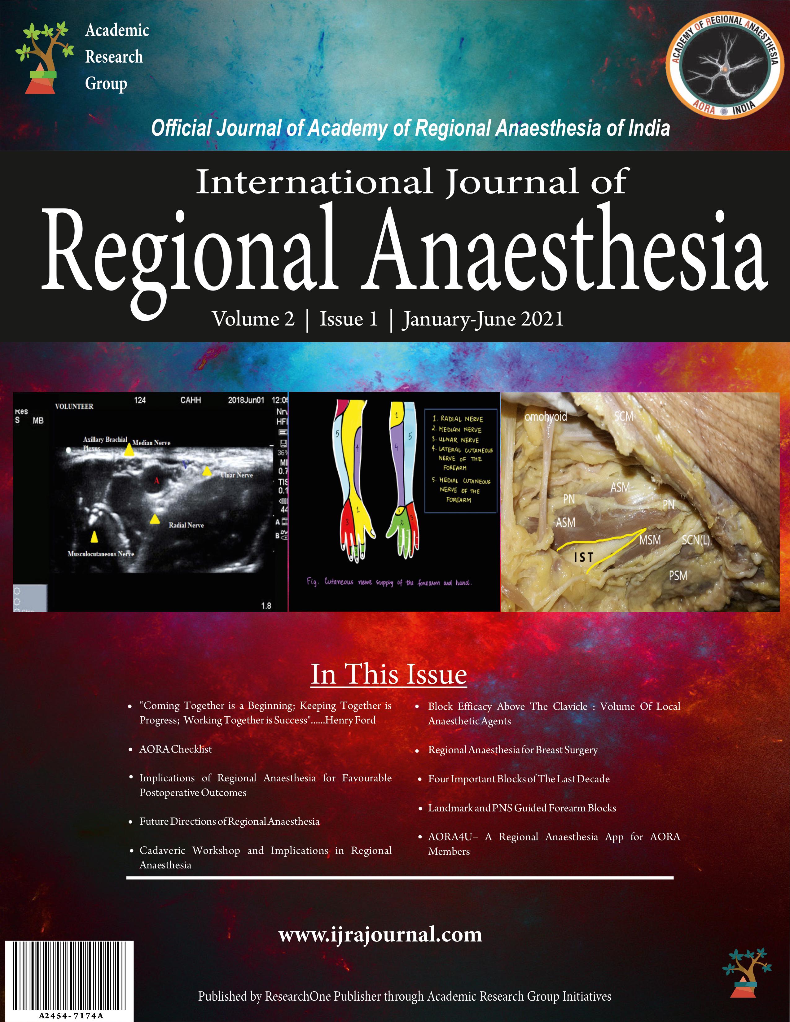
IJRA Cover Page January-June 2021 Vol 2 Issue 1
Acknowledgment: Cover Page Photos.
Dr. Ramkumar Mirle, Dr. Sajana Mukundan & M S Ramaiah Advanced Learning Centre, Bangalore, Karnataka, India. (LHS Figure)
Dr. Surajit Giri & Ms. Shreya Nandini Giri (Class IX, Delhi Public School, Nazira, Sivsagar, Assam.) (Middle Figure)
Dr. Shivaprakash S, Dr. Sandeep Diwan & Department of Anatomy, JSSMC, Mysore, Karnataka, India. (RHS Figure)